Microscopy Community
The microscopy community, chat, and discussion.
100 SOULS
ALL
microscopy
31 posts100 souls
Meet New People
50,000,000+
DOWNLOADS
The microscopy community, chat, and discussion.
100 SOULS
ALL
microscopy
Meet New People
50,000,000+
DOWNLOADS
Related Posts
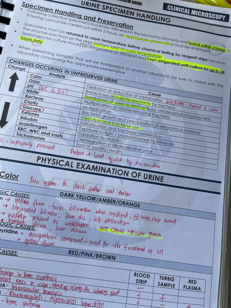
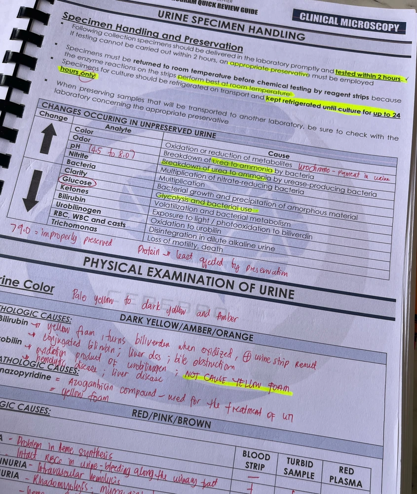
Despite my procrastination (sometimes🙈well, I’m too tired from work), I finished Clinical Chemistry and now welcoming Clinical Microscopy 💩 I’ll give this 5 days max to study 😂 🤞🏻 

What's some of the cool things you get to see at work? For me, it's using polarized light and seeing some fun crystals. In this case our reagent dried out but it looked neat.

Done with clinical microscopy 🥲 I’m so mentally exhausted right now 🥹 I’ll sleep first. Good night~ 
Review season is here! I’m done with clinical chemistry and now in clin microscopy 🥹

I’m very happy to finally return to lab! Even happier to start preparing my first samples for TEM. 👩🏾🔬 🦠Transmission Electron Microscopy (TEM) is a technique to see the tiny details of the materials, sometimes even to the atomic level. It works like an optic microscope,🔬 but instead of… read more

We've talked about some basics on waves, now let's delve into some more practical uses. X-radiation (Xrays) range from 10nm to 10pm in wavelength. This is very useful to us, because that's about the size of atoms all the way down to the size of nuclei. This makes for a wonderful probe at the… read more

I used condensor for oxygenating the samples so the populations will not suffocate, but i do not recommend this, still water is better for them. I think better option is just to fertilise them (with fertiliser for algae) and put them on ambient, not direct light, so the water will not be hot enough… read more

This is literally part of a human cell captured using expansion microscopy. I believe this is a fractal (of?) someone else much larger in the world but Even if it's not I'd super cool to look at.

I took some electron microscopy images and we can’t figure out if they are cavities or bumps on the surface. If you look at it long enough they seem to switch between the two interpretations! Thoughts? Flipped image also included. Thanks! (edited)


Microscopy of water samples i collected from puddles around my house. Seems like a species of Vorticella. (edited)
This is a photo I took of glial cells observed with fluorescence microscopy 🤩 The fluorescent cube is GFP, and it highlights the cell's cytoskeleton. Beautiful

Specifically *Lactifluus volemus var. flavus, aka: "fishy milkcap", and boy are they right. The smell was so strong, I smelled it before I located the fruitbodies themselves. They're a choice edible, but I wasn't 100% sure of identification until the spores were matched. Too late to eat them now. (edited)

A small protozoan that feeds on bacteria. Note the stalk it uses to attach to substrate.


Dunno what this guy is, but I found a ton of them in a saltwater sample. They have a little pointy tail and cilla that seem to feed into it somewhere.

This is bioluminiscent algae from my bought culture at home. I also recorded timelapse of their chloroplast contraction to nucleus.


He did not deserve this.
